Codice criptato: Ipereccitabilità del sistema trigeminale
Codice criptato: Ipereccitabilità del sistema trigeminale
Riassunto

Il soggetto, un uomo di 32 anni affetto da marcato bruxismo notturno e diurno e Dolore Orofacciale (OP) prevalente nelle regioni temporoparietali, con maggiore intensità e frequenza sul lato sinistro della faccia è stato sottoposto al modello diagnostico Masticationpedia che ha decriptato il linguaggio macchina del Sistema Nervoso Centrale in '(Ipereccitabilità' del Sistema Nervoso Centrale con particolare riferimento all'area trigeminale mesencefali. Questa 'Ipereccitabilità' è stata verificata attraverso un metodo elettrofisiologico chiamato ' Ciclo di recupero del Riflesso Inibitorio Masseterino' che evidenziò una esagerato recupero del periodo silente evocato dal secondo stimolo elettrico denominato ' Stimolo test'. Questa condizione neurofisiologica indusse il medico a richiedere una Risonanza Magnetica dell'encefalo che referto un 'Cavernosa Pineale. In conclusione il 'Bruxismo' è una forma di instabilità dell'eccitabilità neurale di tipo funzionale e/od organica, quindi, di non esclusiva pertinenza odontoiatrica. Il perdurare del fenomeno, l'intensificarsi e lo ostinarsi nella gestione dello stesso con trattamenti odontoiatrici senza indagare più dettagliatamente dello 'Stato' di sistema potrebbe essere grave e di prognosi infausta.
Introduzione
Siamo, dunque, giunti nella sezione della Rete Neurale Cognitiva' abbreviata in 'RNC' presentato per la diagnostica del caso della nostra 'Mary Poppins' nel capitolo 'Encrypted code: Ephaptic transmission' e che riproporremo come modello diagnostico per abituare il lettore alla procedura, semplice, intuitiva ma essenziale nei casi clinici di complessa diagnosi come il nostro paziente 'Bruxer'. Il nostro punto di partenza, perciò, è il punto di arrivo della fase precedente alla 'RNC' e cioè la fase discriminatoria dei contesti ( demarcatore di coerenza ). Il basso peso diagnostico derivato dalle asserzioni neurologiche , infatti, é riferito soltanto ad una modesta differenza in ampiezza del jaw jerk. Anche in questo caso la Rete neurale Cognitiva (CNN) ci può venire in aiuto per focalizzare il codice del linguaggio macchina e decriptarlo. Seguiamo, quindi, lo stesso iter già descritto ampiamente nel capitolo 'Encrypted code: Ephaptic transmission' ed avremo la seguente risultato:


Bruxism (4398), trigeminal system (29), abnormality ( 5), excitability ( 3) The excitability of the trigeminal motor system in sleep bruxism: a transcranial magnetic stimulation and brainstem reflex study


Sequenze diagnostiche
1° Step: Sequenza CNN
- Coherence Demarcator: Come abbiamo precedentemente descritto, il primo passo è un comando di inizializzazione della network analysis che deriva, appunto, da una precedente elaborazione cognitiva delle asserzioni nel contesto dentale e quello neurologico a cui il ' Coherence Demarcator' ha dato un peso assoluto eliminando, di fatto, il contesto dentale dal processo. Da quanto emerge dalle asserzioni neurologiche lo 'Stato' del Sistema Nervoso Trigeminale appare relativamente asimmetrico in ampiezza per il jaw jerk, dato una media di . Ciò non consente di inserire nel database Pubmed il comando iniziale puramente neurologico come è stato eseguito per la precedente caso clinico della Mary Poppins. Il comando di inizializzazione sarà, perciò, 'Bruxism' che riguarderà ambedue i campioni di dati (odontoiatrico e neurologico).
- 1st loop open: Questo 'Comando di inizializzazione', quindi, viene considerato come input iniziale per il database Pubmed che risponde con 4398 dati clinici e sperimentali a disposizione del clinico. L'apertura della prima vera analisi cognitiva si elabora proprio sull'analisi del primo risultato del 'CNN' corrispondente a 'Bruxism'. In questa fase, visto la negatività dei referto odomtiatrico e la minima positività del contesto neurologico bisognerebbe individuare una componente neurologico per cui si aggiunge 'Trigeminale system' come 1° loop open.
- 2st loop open: Il processo continua concentrandosi in modo sempre più dettagliato sulle parole chiave che corrispondono ai nostri dati di risultato sul contesto specifico trigeminale che dovremmo completare con un termine riguardante la 'Abnormality' . Questo termine eseguirà il 2° loop open con 5 articoli specifici. A questo punto cognitivamente bisognerebbe fare lo sforzo di valutare tutti i 5 articoli per poter estrapolare qualche indicazione clinica o di laboratorio necessaria alla decriptazione del codice di linguaggio macchina del Sistema Nervoso Centrale. Dalla valutazione degli articoli è emerso un fenomeno possibilmente presente in alcuni casi di bruxismo quello di una alterata eccitabilità del sistema trigeminale. Si è, perciò, inserito nella rete il termine di ‘Excitability' al 3° loop open.
- 3st loop open: In questa fase il dato 'Excitability' ha restituito 3 articoli molto significativi che evidenziano come il livello di eccitabilità del SNC trigeminale si possa testare attraverso una tecnica elettrofisiologica chiamata 'Recovery cycle' del riflesso inibitorio masseterinoe siglato in rcMIR Ovviamente, nel caso in questione, la chiusura del loop della rete è stata fatta sul primo articolo in cui si parla di questa metodologia ( l'ultimo articolo in ordine temporale è stato il nostro). Da questo articolo 'The excitability of the trigeminal motor system in sleep bruxism: a transcranial magnetic stimulation and brainstem reflex study' si evince che nei pazienti che hanno riferito segni e sintomi indicativi di bruxismo notturno (SB), vi é un'eccitabilità anormale delle vie motorie trigeminali. Questa aumentata eccitabilità potrebbe derivare da un'alterata modulazione dei circuiti inibitori del tronco encefalico e non da meccanismi corticali alterati. I risultati supportano l'idea che il bruxismo sia principalmente mediato centralmente e che coinvolga strutture sottocorticali.


2° Step: Ciclo di recupero del Riflesso Massetere Inibitorio
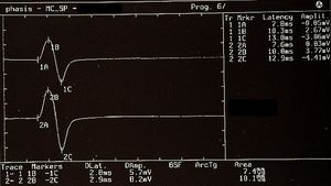
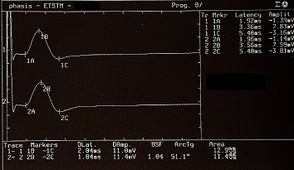
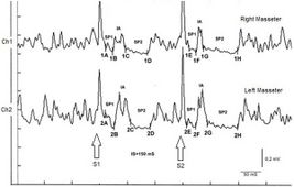
Il ciclo di recupero del Riflesso Massetere Inibitorio (rcMIR) è stato studiato generando coppie di stimoli con caratteristiche identiche, erogati per via percutanea con uno stimolatore elettrico bipolare posizionato sul volto del paziente nella zona del nervo mentoniero. La stimolazione è stata prodotta utilizzando impulsi elettrici ad onda quadra, in grado di evocare un riflesso inibitorio ben definito composto da due periodi silenti (SP), denominati SP1 e SP2, separati da un intervallo di recupero dell'attività EMG “Interposed Activity” (IA). Il primo stimolo (S1) è stato considerato come stimolo condizionante e il secondo (S2) come stimolo test. L'intervallo inter-stimolo tra S1 e S2 è stato impostato a 150 ms.
Al soggetto è stato chiesto di stringere i denti per produrre la massima attività EMG e di mantenere la contrazione per almeno 3 s, con l'aiuto di feedback visivi e audio. Dopo 60 sec di riposo, il soggetto ha ripetuto la contrazione per 10 volte. Il segnale EMG è stato registrato in modalità direttamente rettificata e mediata. Il posizionamento degli elettrodi di registrazione era lo stesso utilizzato per registrare il jaw jerk ed i parametri del preamplificatore sono settati ad una larghezza della finestra temporale di 500 ms, 200 mV per divisione e una larghezza di banda del filtro di 50-1 kHz. Le latenze e le durate degli SP e dell'IA (Figura 1) sono state calcolate come segue:
- Per semplificare l'esame, la rcMIR è stata evocata stimolando elettricamente solo il lato sinistro. Le risposte EMG corrispondono ai tracciati EMG del massetere destro (Ch1) e del massetere sinistro (Ch2). Così, sulle tracce, ogni marcatore indica il numero del canale, mentre le lettere indicano le sequenze delle latenze.
- Lo stimolo S1 suddivide l'acquisizione in pre e post analisi e genera gli SP e l'IA.
- Lo stimolo S2 erogato a 150 ms dal S1, chiamato interstimolo (IS), evoca la seconda sequenza di SP e l'IA.
- Gli SP di S1 e S2 sono determinati automaticamente dal software che posiziona i marker sul primo e sull'ultimo valore minimo elaborato sulle tracce per la generazione di SP1 e SP2, e ne calcola contestualmente la durata. La durata IA è calcolata tra l'ultimo valore minimo di SP1 e il primo valore minimo di SP2.
Nel soggetto testato lo stimolo S2 era in grado di evocare entrambi gli SP, mentre in un soggetto normale lo stimolo S2 di norma è in grado di evocare solo lo SP1 o al massimo un SP2 di durata ridotta. Come mostrato in Tabella 2, la durata dello SP1 evocato da S2 è risultata molto stabile, senza differenze significative nella durata dell'SP1 generato da S1 (Δ= -1ms per Ch1 e Δ= -2 ms per Ch2) mentre l'SP2 evocato da S2 sul massetere destro e sinistro ( 61 ms e 54 ms, rispettivamente) era più lungo di quello evocato da S1 (39 ms e 35 ms, rispettivamente). Le differenze sono state di +22 ms per il Ch1 (massetere destro) e di +19 ms per il Ch2 (mastetere sinistro). Di conseguenza, la durata dell'IA ha mostrato chiare differenze tra S2 e S1. La durata dell'IA evocato da S2 è stata di 12 ms vs. 23 ms dello stimolo S1 per il massetere destro (Ch1) e di 17 ms vs 30 ms di S1 per il massetere sinistro (Ch2) con una differenza tra le risposte evocate da S2 meno S1 rispettivamente di -11 ms e -13 ms.
| Tabella 1 | ||||||
|---|---|---|---|---|---|---|
| Description of the positioning and measurements of the markers
for the recovery cycle of the Masseter Inhibitory Reflex ( rc MIR) |
||||||
| S1 | S2 | |||||
| EMG
traces |
Markers | Onset latency
S1 (msec) |
Markers | Onset latency
S1 (msec) |
||
| 1A | 11 | 13 | 1E | 12 | 12 | |
| Ch1
Right masseter muscle |
1B | 24 | 1F | 24 | ||
| 1C | 47 | 39 | 1G | 37 | 61 | |
| 1D | 86 | 1H | 98 | |||
| 2A | 10 | 16 | 2E | 13 | 14 | |
| Ch2
Left masseter muscle |
2B | 26 | 2F | 27 | ||
| 2C | 56 | 35 | 2G | 44 | 54 | |
| 2D | 91 | 2H | 98 | |||


3° Step: MNR encefalo
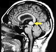
La risonanza magnetica del cervello, utilizzando le sequenze Turbo Spin Echo, Fluid Attenuated Inversion Recovery e Gradient Echo, è stata condotta prima e dopo la somministrazione endovenosa del mezzo di contrasto. I risultati hanno mostrato la presenza di un'area tondeggiante di circa 1,5 cm di diametro situata in prossimità della cisterna quadrigeminale a livello della ghiandola pineale. C'era anche una leggera dilatazione del sistema ventricolare sopratentoriale, che appariva in asse ed era più evidente in prossimità delle corna temporali, con un bordo periventricolare con un fenomeno di assorbimento di liquidi transependimale,[1] caratteristiche che suggerivano una diagnosi provvisoria di cavernoma pineale. (Figura 2 e 3)
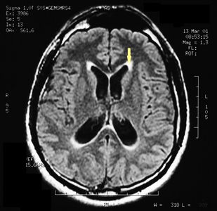
Figure 2: Si può vedere il bordo periventricolare, che indica l'assorbimento transpendimale di liquido (freccia).
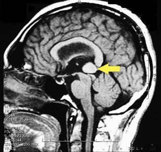
Figure 3: Sul piano sagittale della RM dell'encefalo con mezzo di contrasto (gadolinio) si vede l'esteso cavernoma (freccia).


Considerazioni conclusive
Come possiamo notare dal 'Visual Cognitive gallery', il contesto neurologico si arricchisce del contributo derivante dalla decriptazione del linguaggio macchina attraverso il test del rcMIR ( ) per chiudere definitivamente la diagnosi con il referto della MR ( ). Il modello diagnostico Masticationpedia non solo è di supporto al medico nelle diagnosi complesse ma soprattutto è un elemento di implementazione della nostra conoscenza di base


Sarà utile rappresentare tale interpretazione cognitiva correlandola alle immagini delle asserzioni del contesto neurologico.
- Visual Cognitive gallery
Jaw jerk: As a first step, the assertion that the specific phase highlights an anomaly, even if of minor importance, such as amplitude asymmetry, should always be considered, but the absolute value of the amplitudes on each side should also be noted at the same time. Note in the lower section of the window that the right masseter shows an amplitude of 5 mV while the left one of 8.50 mV. The question to ask is: What is the population mean jaw jerk amplitude? This question is of essential importance because it allows us to understand whether this asymmetry is primarily functional or organic and secondly to quantify its absolute response, but with respect to what? Riflesso mandibolare: Come primo passo, va sempre considerata l'asserzione che videnzi un'anomalia, anche se di minore importanza, come l'asimmetria delle ampiezze, ma va annotato anche il valore assoluto delle ampiezze su ciascun lato. Si noti nella parte inferiore della finestra che il massetere destro mostra un'ampiezza di 5 mV mentre quello sinistro di 8,50 mV. La domanda da porsi è: qual è l'ampiezza media dello riflesso mandibolare nella popolazione? Questa domanda è di fondamentale importanza perché ci permette di capire se questa asimmetria è in primo luogo funzionale od organica e in secondo luogo di quantificarne la risposta assoluta, ma rispetto a cosa?
Trigeminale Root-MEPs: To the motor evoked response of the trigeminal roots recorded on the masseter muscles called bRoot-MEPs.[2][3] This neurophysiological response indicates the anatomical component of the trigeminal motor system of the individual being examined and contextually the absolute amplitude value which, obviously, would correspond to the neuronal energy evoked by the depolarization of all the trigeminal motor fibres contained in the motor root. This very important datum determines the absolute value to which other reflections such as jaw jerk and lateral symmetry refer. Already from these first two questions, we can conclude that the average amplitude of the Root-MEPs we are faced with a datum far beyond the limit, namely which represents a sort of hyper-excitability of the midbrain response.
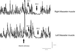
Masseteric Silent Period: Doubt could arise about being in a situation of degenerative and/or demyelinating neuropathy but the test of the electric masseter silent period annuls this hypothesis as both the latency and the duration of the first and second silent period (ES1 and ES2) are symmetrical. Also, the interposed activity of reactivation of the motor units which divides the two silent periods results in symmetry in the integral area between the sides. This data can only highlight hyperexcitability of the trigeminal system by coupling a second electrical stimulus following the first and evoking what has been described, i.e. the recovery cycle of the Inhibitory Masseter Reflex (rcRIM)
Recovery Cycle of MIR: The recovery cycle of the crRIM masseteric inhibitory reflex must be conceptually coupled to the exaggerated amplitude of the jaw jerk because it indicates a condition of hyperexcitability which certainly involves the mesencephalic nuclei with increased excitability of the motoneurons and of the polysynaptic circuitry responsible for inhibition of the motor neuron of the masseter silent period. Take into account the value of the cognitive process of the neural network which has identified the decryption key of neural hyperexcitability in the crRIM. Only with the jaw jerk exam, we would never reached the decryption until the patient's symptoms had worsened enough to require an MR but we all know that this eventuality could be delayed and fatal. Logically, the diagnosis following this process could have been made even 10 years earlier because a sort of attenuated hyperexcitability would certainly have already been present such as to make the physician suspicious.
MR imaging of Pineal Cavernoma: The conclusion of the process as already described indicates that a Cavernosa Pineal is very difficult to attack due to its anatomical position. The patient was referred to a specialist center in neurosurgery in Verona by Prof. Albino Bricolo who succeeded in eliminating the vascular malformation through endoscopic surgery and giving our dear patient 'Bruxer' a dignified life without a dental bite plane.


|
| |||||||


Discussion
The main aim of this study was to electrophysiologically document hyperexcitability of the trigeminal nervous system in a patient affected by pineal cavernoma with pronounced symptoms of OP and bruxism, and who was resistant to any pharmacological or odontological treatment.
We found evidence of activation and peripheral sensitization of the nociceptive fibers, the primary and secondary nociceptive neurons in the CNS, and the endogenous pain control systems, including both the inhibitory and facilitatory processes in our subject.
The concentration of extracellular glutamate in 13 patients affected by cavernous angioma[4] was reported to be increased in comparison with physiological concentrations. High levels of glutamate can cause negative effects on the brain through excitotoxic mechanisms, including degeneration of the superficial layer of the retina in a mouse after repeated administration of glutamate, termed “glutamate excitotoxicity”,[5] resulting from NMDA receptor hyperactivation .[6] In a study in which the trigeminal ganglion neurons were exposed to KCl, the calculated release of glutamate was 10 times greater than the basal level.[7] Further, a significant reduction in the release of potassium-induced glutamate was observed with addition of ω-agatoxin TK, a powerful P/Q calcium channel blocker, while the N-type calcium channel blocker ω-Cgtx conotoxin had a similar effect .[8] Nimodipine, an L-type calcium channel blocker, was also found to reduce the amount of potassium-induced glutamate release.[9] These studies suggest that the P/Q-, N-, and L-type calcium channels each mediate a significant fraction of depolarization-associated glutamate release.
Glutamate release is obviously a much broader and more complex phenomenon. NMDA, kainate, and AMPA ionotrophic receptors, and the metabotropic glutamate receptors, have been found in the superficial lamina of the trigeminal nucleus caudalis in mice.[10] NMDA and AMPA receptor antagonists can block the transmission of the nociceptive trigeminal-vascular signals [11][12] and reduce the high level of c-fos observed in the trigeminal nucleus caudalis following cisternal injection of capsaicin.[13] Furthermore, micro-injections of ω-agatoxin into the ventrolateral area of the periaqueductal gray cause a facilitatory response of nociceptive activity in the trigeminal nucleus caudalis (TNC) activated by tonic electrical stimulation of the supratentorial parietal dura, adjacent to the middle meningeal artery.[14] This response can occur through antinociceptive and/or pronociceptive effects, because the presence of P/Q-type calcium channels is required at the synaptic level for the presynaptic action potentials to couple with the neurotransmitter release processes.[15] Of note, the pre-synaptic afferents in the PAG are positioned on GABAergic inhibitory interneurons and on descending projection neurons. Therefore, the facilitatory effect may be explained by an increased release of GABA, which would indirectly disinhibit the dorsal horn neurons, or by a direct pronociceptive mechanism.[16] These experimental results provide further understanding of the clinical manifestations of pain and central nervous system hyperexcitability found in cases of cerebral cavernous malformations.
Indeed, a blink reflex study on a 38-year-old patient with right hemicranial symptoms associated with a pontine cavernoma affecting the nucleus raphes magnus area revealed a reduction of the pain threshold and a persistent facilitation of the R2 response, with an onset latency difference of 4.4 ms less in the side displaying the symptoms [8]. This confirms a regulatory role for release of neurotransmitters by the nucleus raphes magnus, which exhibits a descending inhibitory control on the TNC [17]and on the entire antinociceptive mesencephalic complex.[18] Our results suggest a hyperexcitability of the trigeminal nervous system in our subject, as follows. First, we evoked a direct response of the trigeminal motor system (bR-MEPs) to provide a value for reference and for amplitude symmetry, as the direct response of the trigeminal motor branch was not affected by any conditioning. A comparison between the jaw jerk responses versus the ipsilateral responses of the R-MEPs showed a much higher amplitude ratio than in normal subjects [19] (Table 1). Therefore, these data indicate the presence of hyperexcitability of the trigeminal system.
The facilitatory effect on the masseter reflex could be indirect. The highest concentration of premotoneurons in the orofacial motor nuclei is found in the bulbar and pontine reticular formations adjacent to the motor nuclei themselves, where these are GABAergic, glycinergic, and glutamatergic-type premotoneurons.[20] In addition, the significant increase of the SP2 recovery cycle from S2 compared with the response from S1 (Table 2) corroborates the hypothesis of hyperexcitability of the trigeminal system. In an in vitro study performed on encephalic slices,[21] intracellular recording of interneurons of the peritrigeminal area (PeriV) surrounding the trigeminal motor nucleus (NVmt) and of the parvocellular reticular formation (PCRt) demonstrated that electrical stimulation of the adjacent areas could evoke both excitatory postsynaptic potentials (EPSPs) and inhibitory postsynaptic potentials (IPSPs). All the EPSPs induced by stimulation of the PeriV, PCRt, and NVmt were shown to be sensitive to ionotropic glutamate receptor antagonists (DNQX and APV), while the IPSPs were sensitive to the GABA and glycine receptor antagonists, bicuculline and strychnine. The cells of this sample showed a long after-hyperpolarization (AHP).
In an electrophysiological study that analyzed a population of neurons and interneurons in the NVmt,[22] three types of AHP were seen: fast, slow, and biphasic. The majority of the motoneurons had a fast AHP (fAHP), whereas most of the interneurons had a slow AHP. The basic properties of these interneurons are similar to the previously described “last-order pre-motoneurons” in the PeriV,[23] suggesting that the interneurons in the NVmt are part of an interneuronal matrix surrounding the NVmt in which the motoneurons are inserted. In this last study, the authors describe the possibility, although rare, of interneurons also having an fAHP.
In our study, the increased duration of the SP2 from S2 invades the IA rather than expanding into the EMG reactivation after the silent period. The afferents for the SP2 descend their intra-axial process along the trigeminal spinal tract and connect with a polysynaptic chain of excitatory interneurons located in the reticular formation at the level of the pontocerebellar junction. The last interneuron in the chain is inhibitory, and sends ipsilateral and controlateral collaterals that ascend medially to the right and left spinal trigeminal complex to reach the trigeminal motoneurons.[24] The interneural sensitization in the rcMIR may be linked to a combination of the excitatory effect of glutamate, with a contribution from the intraneuronal fAHP, and to the disinhibition of the inhibitory processes due to the effect of glycine and GABA.
Overall, our data suggest that certain types of OP, at least those of a central origin, and bruxism are caused by a disruption and homeostatic imbalance of cerebral neurobiochemistry, particularly of the excitatory and inhibitory neurotransmitters in the trigeminal nervous system.
This gives rise to the following questions: Is there a correlation between OP and bruxism, and can bruxism be considered a clinical form of orofacial dystonia?
With respect to the correlation, a distinction should be made between central and peripheral OP on the basis of case history and clinical examination. The muscle discomfort of bruxism is mainly a peripheral phenomenon, resulting from muscle hyperfunction leading to destruction of the myofibrils and release of algogenic substances including myoglobin into bloodstream. By contrast, OP radiating to one or more areas of the face correlated with a clear manifestation of nocturnal or diurnal bruxism could be considered a central type disorder. In these cases, trigeminal electrophysical examinations are highly informative, particularly the rcMIR, blink reflex, JJr, and bR-MEPs, for a differential diagnosis between organic-type lesions of the CNS and functional-type diseases such as TMDs.
Thus, although bruxism and central OP can coexist, they are two independent symptoms, which is why many experimental and clinical studies fail to reach unequivocal conclusions.[25]
It is also possible that bruxism may be a clinical form of dystonia. Our data indicate that bruxism may be a clinical manifestation linked to a CNS neurotransmitter imbalance, and therefore should be considered a subclinical condition of orofacial dystonia or dystonic syndrome. Nevertheless, this phenomenon also appears in a transitory form in children and is resolved with the eruption of mixed dentition.[26][27]
Many studies and diagnostic research protocols, including the Research Diagnostic Criteria (RDC), continue to appear in the field of OP and TMDs, although clear consensus has not yet been reached among the international scientific community.[28] The RDC should consider the patient as affected by a painful syndrome, and should tend towards the definition of a differential diagnosis between organic and/or functional pathologies.[29]
- ↑ Peter H Yang, Alison Almgren-Bell, Hongjie Gu, Anna V Dowling, Sangami Pugazenthi, Kimberly Mackey, Esther B Dupépé, Jennifer M Strahle. Etiology- and region-specific characteristics of transependymal cerebrospinal fluid flow. J Neurosurg Pediatr. 2022 Aug 12;1-11. doi: 10.3171/2022.7.PEDS2246. Online ahead of print.
- ↑ Frisardi G. The use of transcranial stimulation in the fabrication of an occlusal splint. J Prosthet Dent, 1992, DOI: 10.1016/0022-3913(92)90345-b
- ↑ G. Frisardi 1, P. Ravazzani, G. Tognola, F Grandori. Electric versus magnetic transcranial stimulation of the trigeminal system in healthy subjects. Clinical applications in gnathology. J Oral Rehabil.1997 Dec;24(12):920-8. doi: 10.1046/j.1365-2842.1997.00577.x.
- ↑ von Essen C, Rydenhag B, Nystrom B, Mozzi R, van Gelder N, Hamberger A. High levels of glycine and serine as a cause of the seizure symptoms of cavernous angiomas? J Neurochem. 1996;67(1):260–264. [PubMed] [Google Scholar]
- ↑ Lau A, Tymianski M. Glutamate receptors, neurotoxicity and neurodegeneration. Pflugers Arch. 2010;460(2):525–542. doi: 10.1007/s00424-010-0809-1. [PubMed] [CrossRef] [Google Scholar]
- ↑ Meldrum B, Garthwaite J. Excitatory amino acid neurotoxicity and neurodegenerative disease. Trends Pharmacol Sci. 1990;11(9):379–387. doi: 10.1016/0165-6147(90)90184-A. [PubMed] [CrossRef] [Google Scholar]
- ↑ Xiao Y, Richter JA, Hurley JH. Release of glutamate and CGRP from trigeminal ganglion neurons: role of calcium channels and 5-HT1 receptor signaling. Mol Pain. 2008;4:12. doi: 10.1186/1744-8069-4-12. [PMC free article] [PubMed] [CrossRef] [Google Scholar]
- ↑ McCleskey EW, Fox AP, Feldman DH, Cruz LJ, Olivera BM, Tsien RW, Yoshikami D. Omega-conotoxin: direct and persistent blockade of specific types of calcium channels in neurons but not muscle. Proc Natl Acad Sci U S A. 1987;84(12):4327–4331. doi: 10.1073/pnas.84.12.4327. [PMC free article] [PubMed] [CrossRef] [Google Scholar]
- ↑ Hockerman GH, Johnson BD, Abbott MR, Scheuer T, Catterall WA. Molecular determinants of high affinity phenylalkylamine block of L-type calcium channels in transmembrane segment IIIS6 and the pore region of the alpha1 subunit. J Biol Chem. 1997;272(30):18759–18765. doi: 10.1074/jbc.272.30.18759. [PubMed] [CrossRef] [Google Scholar]
- ↑ Tallaksen-Greene SJ, Young AB, Penney JB, Beitz AJ. Excitatory amino acid binding sites in the trigeminal principal sensory and spinal trigeminal nuclei of the rat. Neurosci Let. 1992;141(1):79–83. doi: 10.1016/0304-3940(92)90339-9. [PubMed] [CrossRef] [Google Scholar]
- ↑ Storer RJ, Goadsby PJ. Trigeminovascular nociceptive transmission involves N-methyl-D-aspartate and non-N-methyl-D-aspartate glutamate receptors. Neuroscience. 1999;90(4):1371–1376. doi: 10.1016/S0306-4522(98)00536-3. [PubMed] [CrossRef] [Google Scholar]
- ↑ Goadsby PJ, Classey JD. Glutamatergic transmission in the trigeminal nucleus assessed with local blood flow. Brain Res. 2000;875(1–2):119–124. [PubMed] [Google Scholar]
- ↑ Waeber C, Moskowitz MA, Cutrer FM, Sanchez Del Rio M, Mitsikostas DD. The NMDA receptor antagonist MK-801 reduces capsaicin-induced c-fos expression within rat trigeminal nucleus caudalis. Pain. 1998;76(1–2):239–248. [PubMed] [Google Scholar]
- ↑ Knight YE, Bartsch T, Kaube H, Goadsby PJ. P/Q-type calcium-channel blockade in the periaqueductal gray facilitates trigeminal nociception: a functional genetic link for migraine? J Neurosci. 2002;22(5):RC213. [PMC free article] [PubMed] [Google Scholar]
- ↑ Dunlap K, Luebke JI, Turner TJ. Exocytotic Ca2+ channels in mammalian central neurons. Trends Neurosci. 1995;18(2):89–98. doi: 10.1016/0166-2236(95)93882-X. [PubMed] [CrossRef] [Google Scholar]
- ↑ Pan ZZ, Williams JT, Osborne PB. Opioid actions on single nucleus raphe magnus neurons from rat and guinea-pig in vitro. J Physiol. 1990;427:519–532. [PMC free article] [PubMed] [Google Scholar]
- ↑ Hentall ID. Interactions between brainstem and trigeminal neurons detected by cross-spectral analysis. Neuroscience. 2000;96(3):601–610. doi: 10.1016/S0306-4522(99)00593-X. [PubMed] [CrossRef] [Google Scholar]
- ↑ Jiang M, Behbehani MM. Physiological characteristics of the projection pathway from the medial preoptic to the nucleus raphe magnus of the rat and its modulation by the periaqueductal gray. Pain. 2001;94(2):139–147. doi: 10.1016/S0304-3959(01)00348-7. [PubMed] [CrossRef] [Google Scholar]
- ↑ Cruccu G, Berardelli A, Inghilleri M, Manfredi M. Functional organization of the trigeminal motor system in man. A neurophysiological study. Brain. 1989;112(5):1333–1350. doi: 10.1093/brain/112.5.1333. [PubMed] [CrossRef] [Google Scholar]
- ↑ Li YQ, Takada M, Kaneko T, Mizuno N. GABAergic and glycinergic neurons projecting to the trigeminal motor nucleus: a double labeling study in the rat. J Comp Neurol. 1996;373(4):498–510. doi: 10.1002/(SICI)1096-9861(19960930)373:4<498::AID-CNE3>3.0.CO;2-X. [PubMed] [CrossRef] [Google Scholar]
- ↑ Bourque MJ, Kolta A. Properties and interconnections of trigeminal interneurons of the lateral pontine reticular formation in the rat. J Neurophys. 2001;86(5):2583–2596. [PubMed] [Google Scholar]
- ↑ McDavid S, Verdier D, Lund JP, Kolta A. Electrical properties of interneurons found within the trigeminal motor nucleus. Eur J Neurosci. 2008;28(6):1136–1145. doi: 10.1111/j.1460-9568.2008.06413.x. [PubMed] [CrossRef] [Google Scholar]
- ↑ Kolta A, Westberg KG, Lund JP. Identification of brainstem interneurons projecting to the trigeminal motor nucleus and adjacent structures in the rabbit. J Chem Neuroanat. 2000;19(3):175–195. doi: 10.1016/S0891-0618(00)00061-2. [PubMed] [CrossRef] [Google Scholar]
- ↑ Ongerboer de Visser BW, Cruccu G, Manfredi M, Koelman JH. Effects of brainstem lesions on the masseter inhibitory reflex. Functional mechanisms of reflex pathways. Brain. 1990;113(3):781–792. doi: 10.1093/brain/113.3.781. [PubMed] [CrossRef] [Google Scholar]
- ↑ Svensson P, Jadidi F, Arima T, Baad-Hansen L, Sessle BJ. Relationships between craniofacial pain and bruxism. J Oral Rehabil. 2008;35(7):524–547. doi: 10.1111/j.1365-2842.2008.01852.x. [PubMed] [CrossRef] [Google Scholar]
- ↑ Watts MW, Tan EK, Jankovic J. Bruxism and cranial-cervical dystonia: is there a relationship? Cranio. 1999;17(3):196–201. [PubMed] [Google Scholar]
- ↑ Monaco A, Ciammella NM, Marci MC, Pirro R, Giannoni M. The anxiety in bruxer child. A case–control study. Minerva Stomatol. 2002;51(6):247–250. [PubMed] [Google Scholar]
- ↑ Lobbezoo F, Visscher CM, Naeije M. Some remarks on the RDC/TMD Validation Project: report of an IADR/Toronto-2008 workshop discussion. J Oral Rehabil. 2010;37(10):779–783. doi: 10.1111/j.1365-2842.2010.02091.x. [PubMed] [CrossRef] [Google Scholar]
- ↑ Frisardi G, Chessa G, Sau G, Frisardi F. Trigeminal electrophysiology: a 2 × 2 matrix model for differential diagnosis between temporomandibular disorders and orofacial pain. BMC Musculoskelet Disord. 2010;11:141. doi: 10.1186/1471-2474-11-141. [PMC free article] [PubMed] [CrossRef] [Google Scholar]